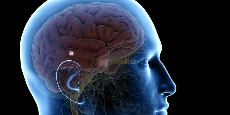
Pineal Tumors
Pineal tumors are a rare occurrence in adults, constituting only 1% of all brain tumors. However, they account for a larger proportion, ranging from 3% to 8%, of intracranial tumors in children, with the typical age at diagnosis being 13 years old.
The most frequently encountered categories of pineal tumors are germ cell tumors (germinoma, teratoma), glial cell tumors (astrocytoma, ependymoma), pineal cell tumors (pineocytoma, pineoblastoma), and miscellaneous tumors (pineal cyst, meningioma). Among all pineal tumors, approximately 10% are benign, an additional 10% are considered relatively benign (low grade), and the remaining 80% are highly malignant.
-
As is the case with many brain tumors, the precise causes of pineal tumors are frequently unclear. Most pineal tumors arise spontaneously and are not associated with genetic factors. The factors that initiate or stimulate abnormal growth are not completely comprehended.
-
Typically, the severity of symptoms will differ depending on various factors such as the size and type of the pineal tumor, whether it disrupts hormone levels or cerebrospinal fluid (CSF) flow, and whether it puts pressure on critical surrounding structures. Symptoms that may be experienced by both children and adults consist of headaches, nausea, fatigue, vision problems, double vision, memory impairment, and seizures. In children, early onset of puberty has been recognized as a significant indicator of a pineal tumor.
-
Magnetic resonance imaging (MRI) is the most important diagnostic tool for tracking the size, shape, and precise location of the pineal tumor, as well as its tendency for growth.
A computed tomography (CT) scan can identify the degree of hydrocephalus (pressure that builds up within the cerebrospinal fluid) and whether the tumor is calcified.
Magnetic Resonance Angiography uses contrast material to provide a look at blood vessels in the body by mapping blood flow and the condition of the blood vessel walls.
-
Due to the remote and challenging-to-access position of the pineal gland, observation (known as the "wait and watch" strategy) is frequently recommended. In situations where treatment is required, the conventional methods entail either one of the open brain procedures or a stereotactic biopsy in conjunction with either radiotherapy, chemotherapy, or both.

